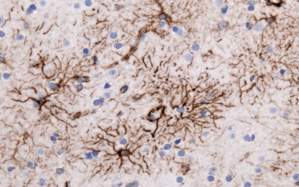
Alexander disease is unusual among neurological disorders because the primary defect is not in neurons, but in astrocytes. The disease is caused by dominant mutations in the gene encoding glial fibrillary acidic protein (GFAP), a structural protein that helps organize the intermediate-filament network inside astrocytes. When mutant GFAP accumulates, it disrupts astrocyte function, promotes formation of Rosenthal fibers, and can damage white matter and other parts of the central nervous system.
GFAP is a structural protein inside astrocytes
GFAP stands for glial fibrillary acidic protein. It belongs to the intermediate-filament family of proteins, which form part of the internal scaffolding that helps cells maintain shape, mechanical stability, and organization.
GFAP is expressed mainly in astrocytes, the star-shaped support cells that regulate many aspects of brain and spinal-cord function. Astrocytes help maintain the extracellular environment, support blood-brain barrier function, regulate neurotransmitter and ion balance, provide metabolic support to neurons, and respond to injury.
GFAP is not required for basic nervous-system development in the same way many neuronal proteins are. Mice lacking GFAP can survive, although they show abnormalities under stress and in specialized astrocyte functions. By contrast, excessive or mutant GFAP can be toxic. That asymmetry is one of the clues that Alexander disease is caused by a toxic gain of function rather than simple loss of GFAP activity.
Alexander disease is a gain-of-function disorder, not a missing-protein disorder
The genetic link was established in 2001, when researchers showed that most cases of Alexander disease are associated with heterozygous mutations in the GFAP gene. The mutations are usually dominant and arise de novo, although familial cases occur.
Unlike disorders caused because a gene no longer works, Alexander disease appears to result from mutant GFAP acquiring harmful properties. GFAP-null mice do not reproduce the characteristic disease, while increasing GFAP expression in experimental models can produce Rosenthal-fiber-like pathology.
This distinction matters therapeutically. If the problem were lack of GFAP, treatment would aim to replace the missing function. Because the problem is toxic accumulation, lowering GFAP becomes a rational strategy.
Rosenthal fibers are the pathological signature of the disease
Rosenthal fibers are cytoplasmic protein aggregates found inside astrocytes. They contain large amounts of GFAP together with small heat-shock proteins such as alphaB-crystallin and other stress-response proteins.
Microscopy studies suggest that Rosenthal fibers can begin as small deposits associated with intermediate-filament bundles and then grow over time. Their presence reflects a broader breakdown in protein handling and astrocyte stress rather than simply acting as inert storage deposits.
In experimental models, astrocytes with heavy GFAP accumulation can become enlarged, lose fine cellular processes, and adopt a reactive phenotype. Those structural changes can interfere with the many support functions astrocytes normally provide to neurons and myelin-producing cells.
The mutation starts in astrocytes, but the effects spread beyond astrocytes
Alexander disease is classified as a primary astrocyte disorder, yet its clinical consequences extend across the nervous system. Patients can develop developmental regression, seizures, spasticity, ataxia, bulbar dysfunction, weakness, autonomic problems, and abnormalities of white matter.
That apparent mismatch is biologically plausible because astrocytes interact closely with neurons, oligodendrocytes, blood vessels, and immune cells. When astrocytes become chronically stressed, secondary effects can alter myelin maintenance, extracellular ion balance, inflammation, and neuronal support.
Our brain health explainer discusses how non-neuronal processes such as vascular and metabolic dysfunction can influence cognition. Alexander disease represents an even more direct example of how disruption of supporting cells can produce severe neurological disease.
Why GFAP levels rise so dramatically
One of the striking features of Alexander disease is that GFAP is not only mutated but also becomes overexpressed. That creates a feed-forward problem: mutant GFAP promotes cellular stress, reactive astrocytes make more GFAP, and increased GFAP creates even more aggregation pressure.
Heat-shock proteins and stress-signaling pathways become activated as cells attempt to manage the abnormal protein load. Older mechanistic work identified activation of pathways such as JNK and sequestration of molecular chaperones into Rosenthal fibers, while more recent studies suggest that some disease-associated GFAP variants undergo abnormal crosslinking and post-translational modification that can further reduce solubility.
The exact initiating events still differ by mutation and remain incompletely understood. What is clear is that total GFAP burden is tightly linked to disease biology.
Animal studies showed that lowering GFAP could reverse established pathology
A key preclinical step came from a 2018 Annals of Neurology study using antisense oligonucleotides in mouse models of Alexander disease. Researchers designed antisense molecules that reduced Gfap messenger RNA and therefore lowered GFAP protein production.
After treatment, GFAP levels fell sharply in the brain and spinal cord. Rosenthal fibers regressed, stress-related molecular markers decreased, microglial activation improved, and treated animals showed improvements in body condition and hippocampal neurogenesis.
The study also found that GFAP protein in cerebrospinal fluid fell with treatment, supporting the idea that CSF GFAP could serve as a pharmacodynamic biomarker.
The investigators described the results as demonstrating the efficacy of antisense suppression for an astrocyte target, providing the mechanistic foundation for clinical development.
Zilganersen turns that mechanism into a human treatment
Zilganersen, marketed as Zanvastro, is a GFAP-directed antisense oligonucleotide. It is designed to bind GFAP messenger RNA and reduce production of the protein before it accumulates further.
FDA approved Zanvastro on September 3, 2026, for pediatric and adult patients with Alexander disease. It is administered by intrathecal injection every three months.
The approval is notable because it directly targets the disease-driving protein rather than treating only symptoms. Our Zanvastro approval article covers the clinical trial and FDA decision in detail.
The randomized study enrolled 49 patients aged 2 years and older, with an additional open-label substudy in 4 children under age 2. FDA reported significantly better walking speed at 61 weeks among treated patients aged 5 years and older who had measurable walking difficulty at baseline. Younger children were evaluated using broader motor-development measures.
Reducing GFAP does not mean eliminating astrocytes
Because GFAP is an astrocyte marker, it can be easy to assume that lowering GFAP might damage or remove astrocytes. That is not what antisense therapy is designed to do.
Zilganersen lowers production of one astrocyte structural protein. The goal is to reduce the pathological excess while leaving astrocytes themselves intact. Preclinical studies were reassuring because substantial GFAP suppression did not simply destroy astrocytes or reproduce Alexander disease.
That strategy is conceptually similar to other precision medicines that lower a toxic protein rather than replacing an absent one. Our rare-disease genetic medicine explainer discusses why genetically defined disorders are particularly suited to this kind of targeted treatment.
What the evidence cannot yet answer
Alexander disease is extremely rare, affecting fewer than 1 in a million people, so both mechanistic studies and clinical trials involve small numbers of patients. Disease severity also varies widely by mutation, age at onset, and affected brain regions.
Many details of the biology remain unresolved, including why some GFAP mutations produce severe early-childhood disease while others cause slower adult-onset disease, how astrocyte dysfunction leads to specific white-matter patterns, and how completely reducing GFAP can halt or reverse established neurological injury.
The pivotal zilganersen study was sponsored by Ionis Pharmaceuticals, the drug’s developer. The randomized design strengthens the clinical evidence, but long-term follow-up will be needed to determine how treatment changes disease trajectory over many years.
It is also too early to know whether reducing GFAP after substantial neurological damage has already occurred can restore lost function or mainly slow further progression.
Alexander disease shows why support cells matter as much as neurons
Alexander disease is a powerful example of how neurological disease can begin outside neurons. A mutation in an astrocyte structural protein can alter protein quality control, stress signaling, white matter, and ultimately the function of the entire nervous system.
That biology also explains why the first disease-modifying treatment targets GFAP itself. The therapeutic logic is unusually direct: if toxic GFAP accumulation drives disease, reducing GFAP production may remove pressure at the source.
References
- Hagemann TL. Alexander disease: models, mechanisms, and medicine. Curr Opin Neurobiol. 2022;72:140-147. DOI: 10.1016/j.conb.2021.10.002.
- Hagemann TL, Powers B, Mazur C, et al. Antisense suppression of glial fibrillary acidic protein as a treatment for Alexander disease. Ann Neurol. 2018;83:27-39. DOI: 10.1002/ana.25118.