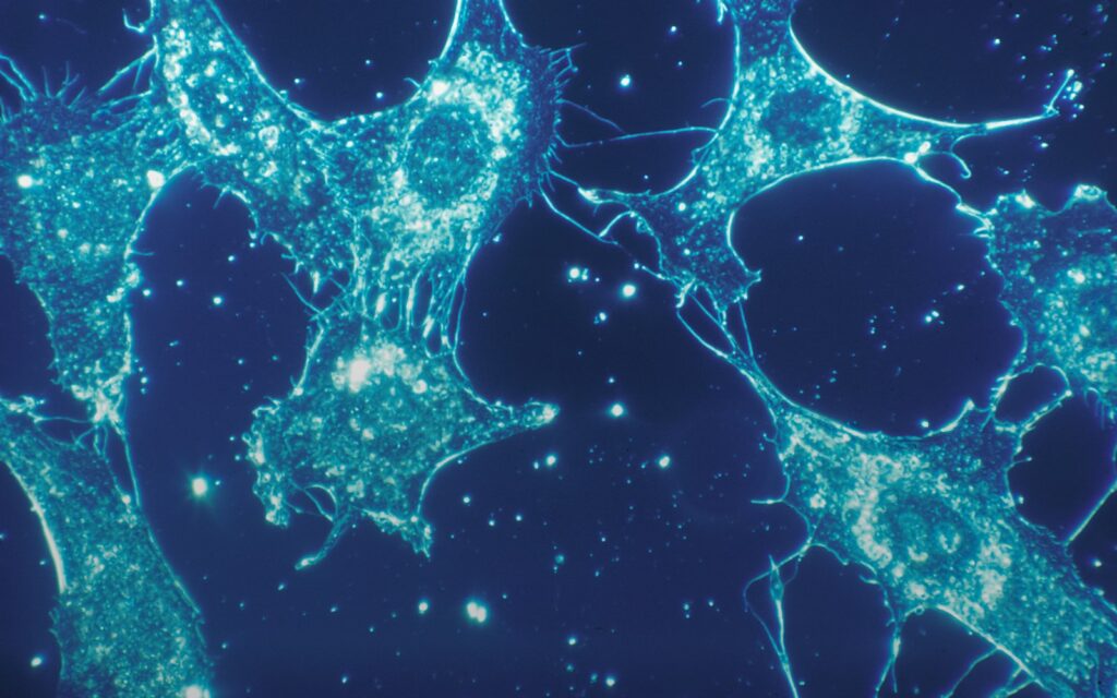
Extracellular vesicles (EVs), the collective term the International Society for Extracellular Vesicles endorsed for lipid-bilayer particles released by cells and unable to replicate, carry protein, DNA, RNA, and lipid cargo between cells across the body. The MISEV2018 guidelines by Théry and colleagues in the Journal of Extracellular Vesicles standardized how the field defines and studies them. A 2020 review by Kalluri and LeBleu in Science synthesized the resulting biology and outlined the emerging clinical applications, from liquid biopsy for cancer to engineered EVs as drug delivery vehicles.
The field spent decades treating EVs as garbage
Cells release membrane-bound particles into the extracellular space. For most of the twentieth century, biologists treated them as debris. That framing broke down when researchers realized the cargo was not random. Cells actively load specific proteins, messenger RNAs, and regulatory small RNAs (including microRNAs) into vesicles before releasing them, and target cells take up those vesicles with a selectivity that suggests coordinated signaling. By the 2010s, EVs were recognized as an intercellular communication channel operating alongside hormones and direct cell contact, capable of modifying gene expression, metabolism, and immune activity in distant tissues.
MISEV2018 sorted out a naming problem
The field’s rapid growth in the 2010s produced a proliferation of names, including exosomes, microvesicles, ectosomes, oncosomes, and apoptotic bodies. Different laboratories used different isolation methods, and terms often referred to populations of overlapping composition. The MISEV2018 guidelines, drafted by Théry and colleagues with over 380 co-authors from the International Society for Extracellular Vesicles, established a common vocabulary. ISEV endorses “extracellular vesicle” as the generic term for particles released by cells that are delimited by a lipid bilayer and cannot replicate. Subtypes are defined operationally by size (small, medium, large), by biogenesis pathway when it can be established, or by the isolation method used. The guidelines also set minimum reporting standards for EV studies to ensure findings could be compared across laboratories.
Two biogenesis pathways produce different subtypes
EVs form through at least two distinct pathways. Exosomes originate inside the cell in structures called multivesicular bodies, which fuse with the plasma membrane to release their contents. Microvesicles (also called ectosomes) bud directly from the plasma membrane. Exosomes are typically 30 to 150 nanometers in diameter (Kalluri and LeBleu report an average of about 100 nanometers); microvesicles are larger and more variable, from about 100 nanometers to a micrometer. The pathways generate vesicles with different cargo profiles because different molecules are sorted to each. In practice, the isolation methods used in most laboratories cannot cleanly separate exosomes from microvesicles, which is one reason the field’s older nomenclature became a source of confusion.
Cargo selection is the mechanistic core
The specificity of EV signaling comes from cargo selection. Cells package particular proteins, RNAs, and lipids into vesicles depending on cell state, environment, and the signaling context. Tumor cells release EVs enriched in tumor-derived proteins and RNAs. Immune cells release EVs that modulate immune responses in target cells. Vesicles from cells under stress carry distinct markers. This selectivity is what makes EVs both a communication channel and a diagnostic opportunity. Blood plasma contains EVs from every organ, and their contents provide a snapshot of the cells that produced them.
Liquid biopsy is the leading clinical application
The most advanced clinical use is in cancer detection and monitoring. Because tumor cells release EVs with a distinctive molecular signature, EVs from a blood sample can carry tumor-specific proteins and RNAs even when the primary tumor is too small to see on imaging. This is one form of “liquid biopsy,” a broad category that also includes cell-free tumor DNA and circulating tumor cells. Diagnostic tests based on EV analysis have been developed for prostate, pancreatic, lung, and other cancers, some now offered clinically. The advantage over conventional imaging is potential sensitivity to earlier and smaller disease; the challenge is distinguishing tumor-derived signal from the substantial background of EVs from healthy tissue.
EVs as drug delivery vehicles are further out
The therapeutic side of the field is earlier stage. EVs have properties that make them attractive as drug carriers: they can cross the blood-brain barrier, they are relatively immune-privileged compared with synthetic nanoparticles, and they can be engineered to display targeting molecules that direct them to specific cell types. Early-phase trials are exploring EVs loaded with anti-inflammatory RNA, chemotherapy agents, or immune-modulating cargo, in indications ranging from oncology to autoimmune disease. None has yet produced a licensed product. The challenges are scale manufacturing, batch consistency, and demonstrating that engineered EVs behave as predicted rather than diluting into the body’s natural EV background.
What the evidence cannot yet answer
Ascribing specific functions to EVs, as the MISEV2018 authors note explicitly, requires reporting details that many published studies have not provided. Much of the literature attributes activity to “exosomes” using isolation methods that also capture other vesicles and non-vesicle contaminants. Whether the specific cargo of EVs functionally alters recipient cells in vivo at physiologically relevant concentrations, and to what extent, is under active investigation. Clinical translation of EV-based diagnostics is proceeding, but standardizing the assays across laboratories and demonstrating that they improve outcomes over existing tests will take large prospective trials. Therapeutic EVs face a harder path, and the field is still working out which cell sources, cargoes, and delivery routes to prioritize.
References
- Théry C, Witwer KW, Aikawa E, et al. Minimal information for studies of extracellular vesicles 2018 (MISEV2018): a position statement of the International Society for Extracellular Vesicles and update of the MISEV2014 guidelines. Journal of Extracellular Vesicles, 2018; 7: 1535750. DOI: 10.1080/20013078.2018.1535750
- Kalluri R, LeBleu VS. The biology, function, and biomedical applications of exosomes. Science, 2020; 367: eaau6977. DOI: 10.1126/science.aau6977
- van Niel G, D’Angelo G, Raposo G. Shedding light on the cell biology of extracellular vesicles. Nature Reviews Molecular Cell Biology, 2018; 19: 213-228. DOI: 10.1038/nrm.2017.125
- International Society for Extracellular Vesicles. About the Society. Available at: isev.org