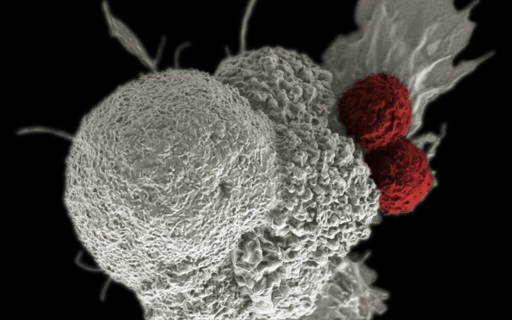
Memory and exhausted T cells follow distinct developmental paths after antigen exposure, with important consequences for cancer immunotherapy, according to research published in Nature and a new multi-omics study published October 21, 2026, in Cell. Experiments in chronically infected mice and human T cell systems show why some T cells retain the ability to renew an immune response while others become progressively constrained by sustained stimulation and altered protein regulation.
Short-lived antigen exposure favors immune memory, while persistent stimulation changes T cell fate
CD8-positive (CD8+) T cells recognize infected or malignant cells through antigen fragments presented on major histocompatibility complex class I molecules. After an acute infection, activated T cells multiply and differentiate into effector cells that can kill targets. As antigen levels fall, much of the expanded population contracts. A smaller pool survives as memory T cells, capable of responding more rapidly to subsequent encounters with the same antigen.
Long-term protection depends on the durability and functional flexibility of these cells. Memory T cells can vary in where they reside, how readily they divide and how quickly they become effectors again. Central-memory, effector-memory and tissue-resident memory populations are not interchangeable.
Cancer presents a different setting. Tumor antigens may remain present for months or years, while cancer cells and surrounding tissues expose T cells to inhibitory signals, limited nutrients and altered metabolic conditions. Prolonged stimulation can direct antigen-specific T cells into an exhaustion-associated differentiation program, rather than the conventional memory pathway.
Exhaustion does not mean that every cell has stopped working. The term describes a heterogeneous family of states, often marked by reduced effector activity, altered proliferative capacity and persistent expression of inhibitory receptors. The distinction helps explain why metabolic signaling linked to T cell exhaustion is an active research target.
Progenitor-exhausted T cells retain renewal capacity without becoming conventional memory cells
A landmark 2016 Nature study by Im and colleagues used mice chronically infected with lymphocytic choriomeningitis virus to distinguish two major virus-specific CD8+ T cell populations. A subset expressing programmed cell death protein 1 (PD-1) and the transcription factor T cell factor 1 (TCF1) could self-renew and generate more differentiated exhausted descendants. It differed from cells already committed to a more terminal exhausted state.
The researchers used cell transfers, transcriptional profiling and PD-1 blockade to establish which populations expanded. The burst of proliferation after blocking PD-1 arose predominantly from the less-differentiated, progenitor-like subset, not from terminally exhausted cells. TCF1 was essential to establishing that progenitor population in the mouse model.
These progenitor-exhausted cells share some features with memory cells, including persistence and self-renewal. They are nevertheless not simply ordinary memory T cells waiting to become active. They arise during chronic antigen exposure and carry a distinct transcriptional and epigenetic identity associated with the exhausted lineage.
Later work has described intermediate exhausted states between progenitors and terminal cells. Different cell populations may also occupy different locations, including lymphoid tissue and tumors. A simple binary division between functional memory and completely inactive exhaustion therefore obscures the biology that matters to treatment.
PD-1 checkpoint blockade can expand a responsive T cell pool without resetting every exhausted cell
Immune checkpoint inhibitors interfere with signals that suppress T cell activity. In selected cancers, blocking the PD-1 pathway has produced substantial and sometimes durable responses. Yet a tumor may contain many cells whose exhausted state is already highly constrained. Removing an inhibitory signal does not necessarily reverse the underlying cell-fate program.
The 2016 mouse study helps explain the difference. It identified a renewal-capable population that could proliferate in response to treatment and produce differentiated descendants. By contrast, terminally exhausted T cells had much less proliferative flexibility. Human tumors are more complex, and the mouse findings cannot by themselves predict which patients will respond to a particular checkpoint inhibitor.
Related questions arise with chimeric antigen receptor (CAR) T cell therapy for solid tumors, where engineered immune cells must remain functional despite persistent antigen exposure and a hostile tumor environment. Maintaining a renewal-capable population may matter as much as maximizing immediate tumor-cell killing.
A 2026 Cell study identifies protein regulation as another layer of exhaustion
Research published in Cell on October 21, 2026, added a different mechanistic perspective. Shuttleworth and colleagues combined RNA sequencing, quantitative protein measurements, protein-turnover analysis and ubiquitin-focused proteomics to examine how T cells change under chronic stimulation.
The investigators studied primary human CD8+ T cells under different stimulation conditions and used additional experimental systems to test selected findings. Their analyses found alterations in protein homeostasis, or proteostasis, including protein synthesis and degradation pathways, alongside changes in metabolism and DNA repair. Some changes at the protein level were not apparent from messenger RNA measurements alone.
One focus was the ubiquitin-proteasome system, which helps control protein stability and turnover. The researchers identified changes in E3 ubiquitin ligases and showed that increasing RNF166 expression improved T cell persistence in experimental systems, including in vivo experiments.
These observations do not establish an RNF166-based therapy for patients. They show that T cell exhaustion cannot be understood entirely by measuring gene transcription or the expression of surface checkpoint receptors. The lifespan and disposal of proteins also affect cellular function.
There is a translational interest in how this might intersect with experimental metabolic reprogramming of engineered T cells. Such approaches may influence persistence, but results from cell cultures and animal models remain separate from clinical efficacy.
What the evidence cannot yet answer
The foundational checkpoint-blockade experiments used a mouse model of chronic viral infection, rather than a randomized treatment trial in patients with cancer. Although those experiments established a mechanism, human tumors contain diverse cell populations and additional signals that can alter T cell behavior.
The newer Cell work included primary human cells but depended heavily on experimentally created stimulation states and mechanistic models. Their molecular patterns may not fully reproduce the immune environment of every tumor. Increased T cell persistence in experimental systems does not prove improved survival, durable tumor clearance or acceptable safety in patients.
Researchers also have not established a single surface marker that perfectly separates beneficial memory cells, progenitor-exhausted cells and terminally exhausted populations across all cancers. Their identities depend on combinations of transcriptional programs, tissue context and stimulation history.
The 2026 Cell authors disclosed relevant commercial interests: some investigators reported biotechnology-company roles, and two researchers were named as co-inventors on a patent application involving RNF166 modulation. These disclosures should be considered when interpreting potential translational applications.
Immunotherapy development increasingly focuses on sustaining useful T cell states
The central challenge is not simply to make T cells more active at one moment. Durable immune control may require preserving cells that can replenish a response, while allowing their descendants to perform immediate tumor killing. Progenitor-exhausted cells and conventional memory cells share some renewal features, but differ in their developmental histories and ability to return to a less-constrained state.
Together, the foundational cell-fate studies and newer protein-turnover findings explain why an effective immunotherapy may depend on more than blocking an inhibitory receptor. It must also operate within the underlying transcriptional, epigenetic and proteostatic programs of the responding cells.
References
- 1. Im SJ, Hashimoto M, Gerner MY, et al. Defining CD8+ T cells that provide the proliferative burst after PD-1 therapy. Nature. 2016;537:417–421. DOI: 10.1038/nature19330.
- 2. Shuttleworth SL, Bauer MA, Province DW, et al. Proteomic atlas of T cell exhaustion defines proteostatic collapse. Cell. Published online October 21, 2026. DOI: 10.1016/j.cell.2026.09.051.